Dr. Gonzalo Vitagliano & Dr. Ríos Pita
Hospital Alemán - Buenos Aires, Argentina
Benefits
✔ Increased understanding of the relationships between the structures involved
✔ Design of the surgical plan
✔ Improved interaction between the doctors involved
✔ Reduction of time in the operating room
✔ Guidance in the surgical approach in the operating room
Clinical case
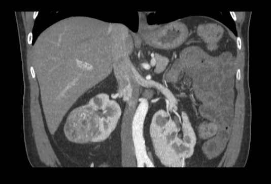
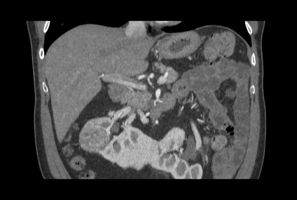
52-year-old man with carcinoma of the right kidney affecting the renal parenchyma and associated vessels.
The patient also presented horseshoe kidney pathology.
3D anatomical model
◾ FDM technology
◾ Material: PLA
◾ Resolution: 0.2 mm
◾ Finish: Multiple colors
Surgical plan and results in the operating room
The tumour was resected laparoscopically.
The 3D model was used to plan the surgical strategy and guide the procedure during the tumour approach. It was especially useful for localising the arteries and interpreting their relationship to the tumour.
"The 3D printed anatomical model perfectly replicated the anatomy of the kidney and the rest of the vasculature to operate more safely and in less time", said Dr. Vitagliano.
Do you want to know more cases in uro-oncology? "Resection of renal tumours with 3D biomodels". At CEMIC, Dr. Nicolás Richards, used 3D printed anatomical models to provide greater patient safety.


