Bronchial tumour: How to avoid resection of a patient's lung?
- MIRAI 3D
- 14 abr 2021
- 1 Min. de lectura
Dr. Alejandro Bertolotti
Hospital Universitario Fundación Favaloro - Buenos Aires, Argentina
Benefits
✔ Increased understanding of anatomy
✔ Redesign of surgical plan
✔ Improved patient-physician interaction
✔ Guidance in the surgical approach in the operating room
Clinical case
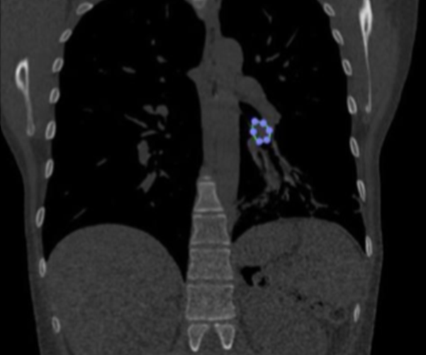
38-year-old female former smoker diagnosed with bronchial carcinoid tumour located in the lower lobe of the left lung.
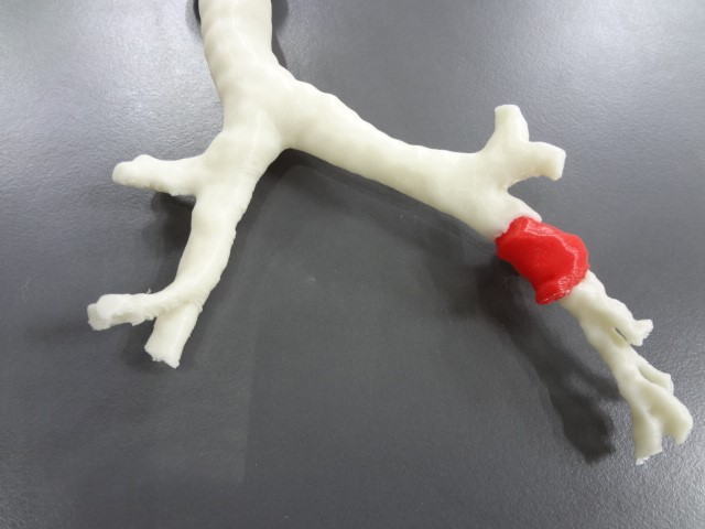
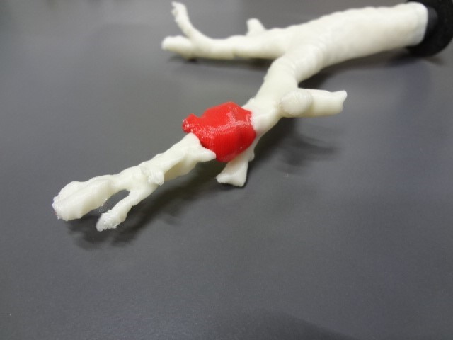
3D anatomical model
◾ Technology: FDM
◾ Material: PLA
◾ Resolución: 0.2 mm
◾ Finish: Two colors
Surgical plan and results in operating room
The first surgical plan, based on the CT images, was a left lower lobectomy.
After having the 3D model of the patient's airways available, the surgeon was able to precisely locate the tumour and noticed that it was lower than he thought. In addition, the three-dimensional model made it easy to identify its boundaries, which allowed him to think of a new plan for the patient's treatment.
Thus, the surgical strategy was changed and a segmental bronchial resection was performed, which involved removing only the tumour while sparing the patient's lower lobe.
Reanastomosis of the bronchial tree was then performed to reconstruct the airway without loss of lung parenchyma.
Testimonial


