Endobronchial Tumour - 3D Biomodelling Assisted Thoracoscopy
- MIRAI 3D
- 16 abr 2021
- 2 Min. de lectura
Dr. Matías Nicolás
Hospital Privado de Comunidad - Mar del Plata, Argentina
Benefits
✔ Increased understanding of the lesion and its environment
✔ Confirming the chosen surgical strategy
✔ Improved safety
✔ Improved patient-physician interaction
Clinical case
In this clinical case, a 78-year-old male patient presented with haemoptysis at the doctor's office. As a result, he was instructed to undergo appropriate tests to evaluate his clinical picture.
An initial CT scan showed a single endobronchial lesion at the level of the middle bronchus. A fibrobronchoscopy was performed and a biopsy was taken which showed that the lesion was an undifferentiated adenocarcinoma type neoplasm.
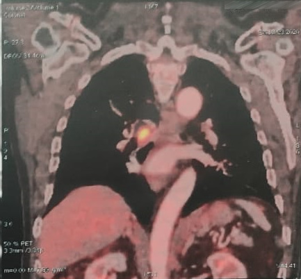
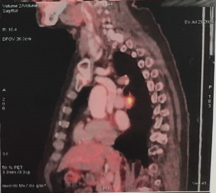
Finally, a PET CT scan showed hypermetabolic activity in the lesion at the level of the middle bronchus with no evidence of distant disease, so it was decided to perform resective surgery.
The tomographic studies were used to make the 3D model requested to evaluate the surgical procedure to be performed.
3D anatomical model
◾ FDM Technology
◾ Material: PLA
◾ Resolution: 0.2 mm
◾ Finish: Three color
Surgical plan and results in operating room
A cuff resection procedure of the bronchus intermedius via uniportal videothoracoscopy (UVATS) was chosen for the surgical intervention.
The 3D model was useful to better understand the location and extent of the tumour and to ensure that the previously selected surgical procedure was appropriate for the patient in question. This resulted in greater safety for Dr. Nicolás when operating.
You may be interested in: "Bronchial tumour: How to avoid resection of a patient's lung". Discover Dr Bertolotti's case, at Fundación Favaloro, where the 3D model played a crucial role in defining the surgical strategy.
In addition, the biomodel was used to explain the procedure to the patient. In this regard, the attending physician said that "the patient and his family felt that the plan for the surgery covered all the possibilities and were surprised to be able to see a life-size image of the tumour".
Video of CBF before and after resection
Do you want to know more applications in thoracic surgery?
Free online course: INNOVATION IN SURGERY.
Enter to www.modelosmedicos.com/webinar to watch on-demand webinars.


